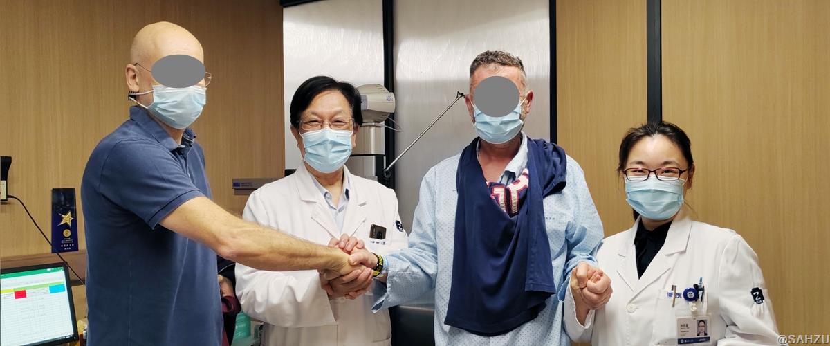
 DR. LIN JIJIAN (2ND FROM LEFT) AND DR. LIN YUCHEN (4TH FROM LEFT) HOLDING HANDS WITH PAOLO (3RD FROM LEFT) AND HIS FRIEND
Recently, an international patient showed up at Dr. LIN Jijian's clinic, looking very much worried. He might need help. Dr. LIN immediately sensed his anxiety.
Sudden Vision Loss on the International Flight
It turns out that Paulo the patient was flying to Hangzhou from Italy for a business trip, when he suddenly felt rapid reduction of eyesight in his right eye with floating black shadows over obscured field of vision, making him very difficult to see.
After an unsettling 13-hour flight, Paolo immediately rushed to a nearby hospital once landing in Hangzhou. He was diagnosed with sudden retinal detachment in his right eye and recommended to SAHZU Eye Center for further and more precise treatment.
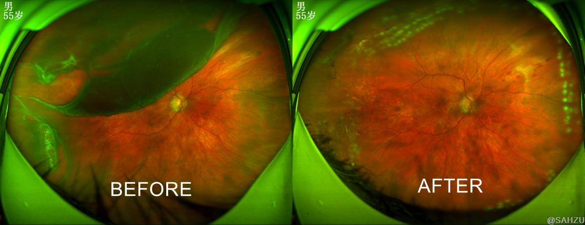
At Dr. LIN's clinic, Paolo was measured -6.75 diopters of myopia of his both eyes. And there were two tears above the half-detached retina of his right eye, causing black shadow over the field of vision and a decimal visual acuity of only 0.1.
Dr. LIN also learned that Paolo once had a retinal tear in his left eye before, which was treated with laser at an Italian hospital to prevent retinal detachment. But unfortunately, his right eye was not spared this time.
Why is retina detached?
The retina is a transparent, film-like, thin layer of tissue located at the back of the eye, close to the inner wall of the eye, and covered with light-sensitive cells and nerve fibers.
If we compare our eyes to a camera, the retina is like the camera's negative, which is responsible for recording the image information and converting it into signals to be transmitted to the brain. If the camera's negative is damaged, no picture can be taken. Similarly, the condition of retinal detachment will deteriorate rapidly, resulting in vision decrease and blockage of the field of vision, until permanent vision loss. Surgery is the only treatment.
There are typically three types of retinal detachment.
Rhegmatogenous retinal detachment happens when there is a tear in the retina.
Tractional retinal detachment happens when retina is pulled away by vitreous body pathologically adhered to retina and torn.
Exudative retinal detachment happens when there is an abnormality in the retina itself or in the surrounding structures leading to leakage of fluid from the blood vessels, and fluid builds up in the subretinal space
Paolo's case is a typical rhegmatogenous retinal detachment. "The patient was 55 years old and had been highly myopic for many years. Myopia lengthens the eye axis and gradually expands the posterior eye wall. As time passes, the retina is prone to degeneration, atrophy, and thinning. High myopia therefore is an key risk factor for retinal detachment. His long-distance flight and exhaustion may also have induced retinal detachment. In addition, aging, eye trauma, diabetic retinopathy and other factors may also lead to retinal detachment." Said Dr. LIN.

3D visualization system-assisted eye surgery
Retinal detachment is a serious blinding disease that progresses rapidly. If left untreated, the detachment will become more extensive, resulting in greater visual impairment and less effective treatment, which may lead to blindness eventually.
Paolo was rushed to the surgery room the next day. Dr. LIN performed 3D high resolution visualization system-assisted vitrectomy and retinal detachment repair. SAHZU Eye Hospital is one of the earliest in China to perform 3D visualization system-assisted eye surgery, and the forerunner in both the volume and surgery skills of vitreoretinal surgery.
During the procedure, Dr. LIN observed on the 3D visualization system that another two tears around the periphery of the retina were forming, and the detachment range was wider than that of the previous day. Dr. Lin performed the vitrectomy + intraocular laser surgery + intraocular silicone oil implantation on the patient’s right eye, and repaired and restored the detached retina.
The surgery went very smoothly. The 3D visualization system is a highly precise surgery system developed in the last three years. Its ultra-large magnification, 4K high-resolution, and high depth of field, create a better three-dimensional visual effect and more detailed display than the traditional microscope, which can realize the fine rendering of the tissue structure during the surgery and the precise positioning of the surgical maneuver. The system could help to reduce the accidental injuries during the surgery and lower the surgical risks.
Paolo had no nausea, vomiting, dizziness, or other complications after the surgery with all vital signs remained good.
The next day after the surgery, Dr. Lin checked on Paolo. The examination showed that the retina had been restored, tears healed well, and dark shadows in front of his right eye completely disappeared. His visual acuity had reached 0.3. Paolo showed his first smile since his admission, gave Dr. Lin a thumbs-up, and said "Grazie”.
In view of Paolo's long flight back to Italy, Dr. Lin did a laser surgery again to prevent retinal detachment in his left eye.
Author: LI JING | Reviewer: CHEN LU | Editor: LI JING | Source: SAHZU EYE CENTER | Date:2023-07-27 | Views:![]()
![]()