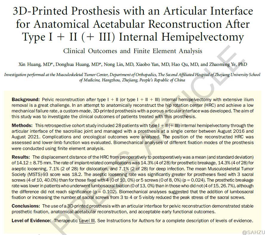
The anatomical complexity of the pelvic region, located near critical blood vessels, nerves, and organs, presents immense challenges for surgical resection and reconstruction of malignant tumors. Tumors in the pelvic zones I + II (+ III) are particularly difficult due to the extensive bone and soft tissue loss following internal hemipelvectomy. Stabilizing the pelvic structure and precisely reconstructing the hip joint in its anatomical position remain significant challenges. Currently, common international approaches include leaving the hemipelvis unconstructed or even performing hemi-pelvic amputations, which result in limited postoperative function.
Chair of SAHZU Orthopedics Prof. YE Zhaoming's team has spent years refining techniques for pelvic tumor resection and reconstruction. To address these clinical challenges, they introduced groundbreaking advancements in both tumor resection and pelvic prosthesis reconstruction. Their research work has been recently published on the Journal of Bone and Joint Surgery (JBJS), the world top journal in the field of orthopedics, titled "3D-Printed Prosthesis with an Articular Interface for Anatomical Acetabular Reconstruction After Type I + II (+ III) Internal Hemipelvectomy: Clinical Outcomes and Finite Element Analysis."
Dr. Huang Xin is the article’s first author, Dr. Huang Donghua co-first author, and Prof. YE Zhaoming the corresponding author.
(Click here to view the full article)

Tumor Resection
Traditional procedures use ultrasonic bone knife, wire or oscillating saws to vertically cut through the ilium or sacrum, compromising the sacroiliac joint surface. This study innovatively severs the ligaments using electrosurgical knife, causing dislocation of the sacroiliac joint. Such procedure preserves the intact sacroiliac joint surface to facilitate subsequent precise reconstruction.
Prosthesis Design
The team developed a 3D-printed, custom-made hemipelvic prosthesis with an articular interface (Figure 1). The porous articular interface was designed to seamlessly fit the articular interface of the sacrum, and the acetabular component is designed to position the hip joint accurately in its anatomical center.

Figure 1 The design diagram and actual image of the prosthesis
Prosthesis Installation
Customized sacral screws in varying numbers, directions, and angles are used to fix the prosthesis, reducing shear stress at the sacroiliac interface and minimizing the risk of loosening and breakage.
Follow-up Results
Most patients reported satisfactory limb function with low rates of prosthesis-related complications, including loosening, breakage, or dislocation, alongside favorable oncological outcomes.
Finite Element Analysis
The study validated these clinical findings with finite element analysis (Figure 2), demonstrating that lumbosacral fixation or using more than three sacral screws significantly reduces the maximum stress on screws, lowering the risk of screw fracture and prosthesis loosening.

Figure 2 Preoperative and postoperative radiological images and schematic of finite element analysis
JBJS is a top-tier, Nature-indexed orthopedic journal, and the official publication of the American Orthopedic Association (AOA). It is recognized as a leading medical journal in the field of orthopedics, holding high impact and prestige.
Founded in 1953 and widely known as the "Cradle of Orthopedic Surgeons in Zhejiang", the Department of Orthopedics at SAHZU is the oldest and largest orthopedic department in Zhejiang Province. It is recognized as a National Clinical Key Specialty and a Provincial Key Discipline. The department has 171 surgeons in total, including 32 chief physicians, and 61 associate chief physicians. With 417 beds, the department serves about 650,000 outpatient visits annually, and over 35,000 surgeries are performed each year.
The department is leading in the treatment of complex and critical orthopedic cases, utilizing advanced surgical techniques from around the world.
The department’s bone tumor center is one of the earliest and largest centers of its kind in China, specializing in comprehensive treatment for malignant tumors, including osteosarcoma, Ewing sarcoma, and chondrosarcoma. The team excels in advanced techniques such as pelvic tumor resection and reconstruction, total vertebral resection for spinal tumors, 3D-printed prosthetic reconstruction, and minimally invasive tumor treatments. Early on, the bone tumor center established a multidisciplinary team (MDT) incorporating oncology, radiology, pathology, and radiation therapy to provide patients with precise, standardized, and personalized care informed by cutting-edge, international standards.
Author: | Reviewer: LI JING | Editor: LI JING | Source:HUANG DONGHUA | Date:2024-11-18 | Views:![]()
![]()